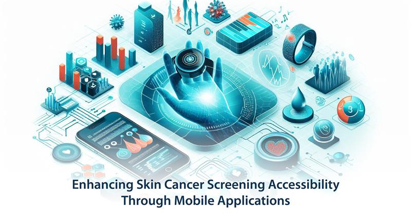
Enhancing Skin Cancer Screening Accessibility Through Mobile Applications

Introduction

Alpha and beta cancers rank as the leading form of malignancy worldwide because they produce fresh cases in millions annually. Public health outcomes strengthen when medical access occurs early enough for lifesaving treatment while expenditures decline. The limitation of prompt screening services mainly targets regions that do not have proper medical infrastructure in remote areas. The physical barriers for patients to visit dermatologists exist because screening appointments traditionally rely on in-person medical visits. The development of mobile technology provides society with an unmatched opportunity to deal with these barriers. Outside medical facilities, healthcare providers now use mobile applications to expand their screening services, which leads to simpler early detection opportunities. The study explores mobile applications as a means for skin cancer screening using technical explanations together with analyses of implementation prospects and present limitations in its approach.

The Importance of Early Skin Cancer Detection

Successful skin cancer treatment becomes more probable when patients detect their condition at its primary stage. A timely diagnosis of skin cancer called melanoma improves the treatment process when the cancer is found early. The delayed diagnosis of skin cancer happens because patients face barriers to accessing dermatologists and healthcare facilities that meet specialized needs. Mobile applications serve as a solution by giving users tools to conduct preliminary skin cancer evaluations and close the gap between healthcare providers and patients.

Role of Mobile Applications in Skin Cancer Screening

Advanced image processing algorithms and machine learning techniques inside mobile applications evaluate skin lesions for the purpose of skin cancer screening. Users can take pictures of unusual skin growths using their smartphone camera function. The applications assess captured images based on their color tones and their size specification, as well as their shape properties. Risk assessment scores included in some applications enable users to evaluate their situation regarding seeking medical advice.

Yearwise Publication Trend on skin cancer

Year Publication Count
2025 9
2024 3625
2023 6045
2022 3521
2021 2710
2020 2184
2019 1492
2018 1081
2017 880
2016 1009
2015 796
Find publication trends on relevant topics

Technologies Powering Mobile Screening Solutions

Image Processing and Analysis

The assessment of skin lesions benefits from advanced image processing techniques which modern mobile applications utilize precisely. These technologies include:

  • Segmentation Algorithms: To isolate the lesion from the surrounding skin.
  • Feature Extraction: To analyze key visual characteristics.
  • Classification Models: To categorize lesions based on risk factors.

Machine Learning and Artificial Intelligence

Mobile screening applications achieve better accuracy levels through their implementation of Artificial Intelligence (AI) and Machine Learning (ML). Accurate systems for risky lesion detection develop through extensive training of ML models from large dermoscopic databases. The analytic models of AI and ML enhance their capabilities through the observation of new data and subsequent analytic processes.

User-Friendly Interfaces

Mobile applications contain user-friendly interfaces for diverse technical abilities to gain accessibility. This application improves user experience by offering guidance for image capture combined with instructional steps that provide immediate feedback to users who perform regular skin checks.

Benefits of Mobile Applications in Skin Cancer Screening

Increased Accessibility

Mobile applications remove geographical limitations to provide people in remote settings with basic screening assessment tools. Mobile applications provide crucial access to health services because these areas lack proper dermatological support.

Cost-Effective Screening

Mobile applications function as a budget-friendly solution that minimizes the requirement of first face-to-face appointments for early skin cancer discovery. Users can do self-examinations when they want which suppresses the demand on healthcare facilities.

Empowering Patients

Through mobile screening applications, users gain the ability to participate actively in their healthcare. The practice of systematic self-inspection helps patients identify conditions early which results in better medical outcomes with higher survival possibilities.

Recent Publications on skin cancer

Challenges and Limitations

Despite their advantages, mobile applications for skin cancer screening face several challenges:

The diagnostic accuracy of AI and ML models requires further development because there are still improvement possibilities.

Acquiring necessary regulatory approvals required for medical applications becomes a demanding procedure that can take a long time to complete.

Reliable outcomes from skin cancer screening systems require users to properly follow procedures when they take images and operate the application.

Future Prospects

Mobile skin cancer screening technology will continue advancing in promising directions. Continuous development in artificial intelligence systems together with expanding training data collections improves medical diagnosis performance. The integration of technology companies with healthcare providers results in platform development that enables fluid communication between patients and doctors. The diagnostic process can become more efficient through telemedicine integration which enables real-time doctor consultations and patient follow-up services within these applications

Conclusion

The innovative nature of mobile applications brings substantial changes to skin cancer screening by implementing better accessibility cost-efficient operation and user-friendly approach. Modern technology and productive healthcare partnership structures will solve existing healthcare challenges. Mobile screening technology functions as the main instrument for minimizing skin cancer cases globally by advancing its adoption and development platform.

References

  1. Giger, M.L., 2018. Machine learning in medical imaging. Journal of the American College of Radiology15(3), pp.512-520.
  2. Masood, A. and Al-Jumaily, A.A., 2013. Computer aided diagnostic support system for skin cancer: a review of techniques and algorithms. International journal of biomedical imaging2013(1), p.323268.
  3. Ramlakhan, K. and Shang, Y., 2011, November. A mobile automated skin lesion classification system. In 2011 IEEE 23rd International Conference on Tools with Artificial Intelligence (pp. 138-141). IEEE.
  4. Rosado, B., Menzies, S., Harbauer, A., Pehamberger, H., Wolff, K., Binder, M. and Kittler, H., 2003. Accuracy of computer diagnosis of melanoma: a quantitative meta-analysis. Archives of Dermatology139(3), pp.361-367.
  5. Gouda, W., Sama, N.U., Al-Waakid, G., Humayun, M. and Jhanjhi, N.Z., 2022, June. Detection of skin cancer based on skin lesion images using deep learning. In Healthcare (Vol. 10, No. 7, p. 1183). MDPI.
  6. Bechelli, S. and Delhommelle, J., 2022. Machine learning and deep learning algorithms for skin cancer classification from dermoscopic images. Bioengineering9(3), p.97.
  7. Zhang, Y. and Wang, C., 2021, March. SIIM-ISIC melanoma classification with DenseNet. In 2021 IEEE 2nd International Conference on Big Data, Artificial Intelligence and Internet of Things Engineering (ICBAIE) (pp. 14-17). IEEE.
  8. Poornima, A., Devi, M.S., Sumithra, M., Bharath, M.V., Sathishkumar, S., Yogesh, K., Upadhyay, S.S. and Sah, N.K., 2021, February. Epoch interrogation for skin cancer detection using convolutional neural network models. In IOP Conference Series: Materials Science and Engineering (Vol. 1074, No. 1, p. 012025). IOP Publishing.
  9. Masood, A. and Al-Jumaily, A.A., 2013. Computer aided diagnostic support system for skin cancer: a review of techniques and algorithms. International journal of biomedical imaging2013(1), p.323268.

Top Experts on “skin cancer

S. Thrun

H-Index: 142
Publication Count: 439
Citation Count: 102290

Affiliation: Department of Computer Science, Stanford University, Stanford, CA, USA.

F. Clavel-Chapelon

H-Index: 135
Publication Count: 713
Citation Count: 66346

Affiliation: Inserm, Centre for Research in Epidemiology and Population Health (CESP), U1018, "Nutrition, Hormones, and Women's Health" Team, Institut Gustave Roussy, F-94805, Villejuif, France.

M. Klagsbrun

H-Index: 108
Publication Count: 327
Citation Count: 43244

Affiliation: Vascular Biology Program, Boston Children's Hospital, Harvard Medical School, Boston, MA 02115, USA; Department of Surgery, Boston Children's Hospital, Harvard Medical School, Boston, MA 02115, USA; Department of Pathology, Boston Children's Hospital, Harvard Medical School, Boston, MA 02115, USA.

J. Cerhan

H-Index: 108
Publication Count: 1034
Citation Count: 46288

Affiliation: Department of Health Sciences Research, Mayo Clinic, Rochester, MN, USA.

C. Garbe

H-Index: 104
Publication Count: 873
Citation Count: 56269

Affiliation: Center for Dermatooncology, Department of Dermatology, University Hospital Tuebingen, Liebermeisterstr. 25, 72076, Tuebingen, Germany.

J. Overgaard

H-Index: 103
Publication Count: 979
Citation Count: 50123

Affiliation: Department of Experimental Clinical Oncology, Aarhus University Hospital, DK-8000 Aarhus C, Denmark.

E. Loftus

H-Index: 103
Publication Count: 979
Citation Count: 46372

Affiliation: Division of Gastroenterology and Hepatology, Mayo Clinic, Rochester, Minnesota.

J. Seddon

H-Index: 98
Publication Count: 378
Citation Count: 36297

Affiliation: Ophthalmic Epidemiology and Genetics Service, New England Eye Center, Tufts Medical Center, Boston, MA, USA, Department of Ophthalmology, Tufts University School of Medicine, Boston, MA, USA and Sackler School of Graduate Biomedical Sciences, Tufts University, Boston, MA, USA

G. Pfeifer

H-Index: 95
Publication Count: 366
Citation Count: 36524

Affiliation: Department of Cancer Biology, Beckman Research Institute, City of Hope, Duarte, CA 91010, USA Department of Diabetes and Metabolic Diseases Research, Beckman Research Institute, City of Hope, Duarte, CA 91010, USA

X. Castellsagué

H-Index: 95
Publication Count: 276
Citation Count: 45899

Affiliation: Institut Catala d'Oncologia (ICO), IDIBELL, CIBERESP, L'Hospitalet de Llobregat, Catalonia, Spain.